Cartilage
Cartilage is a thin, elastic tissue that protects the bone and makes certain that the joint surfaces can slide easily over each other.
There are two types of joint cartilage in the knees:
- Fibrous cartilage: (meniscus), which has tensile strength and can resist pressure.
- Hyaline cartilage: Which covers the surface along which the joints move.
Cartilage will wear over the years, as it has a very limited capacity for self-restoration. The newly formed [healed Scar] tissue will generally consist for a large part of fibrous cartilage of lesser quality than the original hyaline cartilage. As a result, new cracks and tears will form in the cartilage over time.
What is a meniscus?
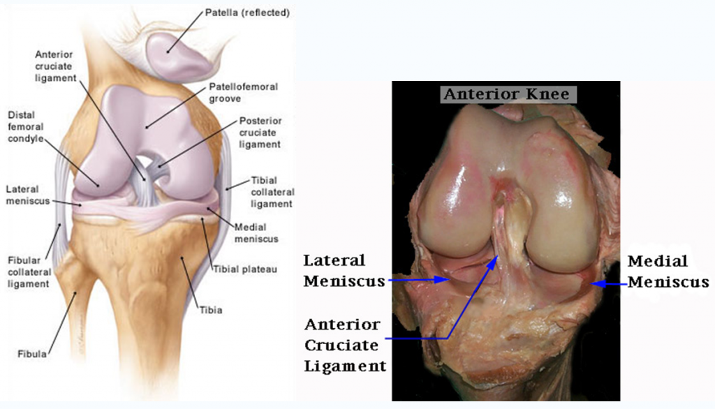
- The menisci (plural of meniscus) are horizontal horse shoe shaped wedges of tissue that exist both on the inside and outside aspects of the knee acting as shock absorbers between the long bones making up the knee.
- Each knee has 2 menisci, one to support each of the rounded ends (condyle) of the femur (thigh bone).
- They are made of smooth white glistening fibrous material, and have a vital function in reducing dissipating shock forces acting on the knee during day to day activity.
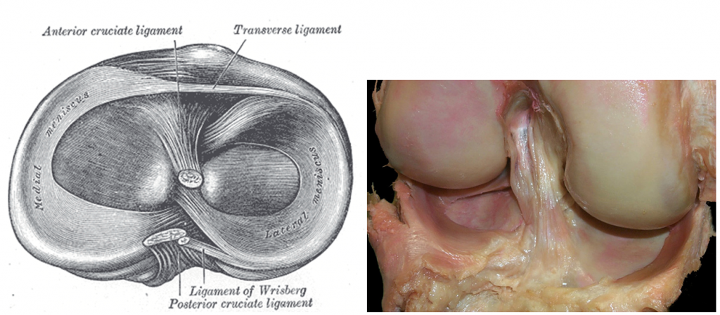
- They are wedge shaped and curved, with the wider part of the wedge forming the outer rim and the inner rim being the sharp surface.
- The majority of the meniscus does not have a blood supply and it is for this reason that healing of these tissues once damaged is very unlikely.
- These two disks, the medial meniscus and the lateral meniscus, consist of connective tissue with extensive collagen fibers containing cartilage-like cells. Strong fibers run along the menisci from one attachment to the other, while weaker radial fibers are interlaced with the former.
- The menisci serve to protect the ends of the bones from rubbing on each other and to effectively deepen the tibial sockets into which the femur attaches.
- They also play a role in shock absorption, and may be cracked, or torn, when the knee is forcefully rotated and/or bent.
- Consequently, people who have damaged their cartilages often ultimately require surgery to excise the torn fragments.
Articular Cartilage:
The Articular Cartilage, also known as the joint lining,(Hyaline cartilage) is a protective layer of tissue located on the ends of the bones which come together in the knee joint. These bones are the femur (thigh bone), tibia (shin bone), and patella (kneecap).
Articular cartilage can be damaged through an injury or gradually deteriorate over time from a variety of factors. When the articular cartilage is damaged or injured, it usually goes through a staged process of softening, flaking, fragmenting, and finally complete loss where the underlying subchondral bone is exposed. This process is commonly known as osteoarthritis.
Damaged or injured articular cartilage has a very limited ability to heal itself. Therefore, once the process of osteoarthritis starts, there is little that the body can do to stop the deterioration.